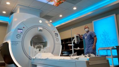
If you have unexplained symptoms in your chest or unexpected findings from a chest X-ray or another imaging study, your physician may order a chest CT scan or MRI scan to help make a diagnosis. These versatile forms of advanced imaging can show organs, soft tissue, blood vessels and abnormalities, such as tumors, in greater detail. As a result, chest CT scans and MRI scans can help physicians diagnose a wide range of diseases and injuries.
Both a chest CT scan and an MRI scan create cross-sectional images of internal structures — think of slices of bread that, together, form a loaf — but in different ways. CT scans produce images using an X-ray beam that rotates around you. MRI scans, on the other hand, use magnets and radio waves. You may need to hold your breath for a short period of time during certain CT scans or MRI scans to ensure you are completely still while images are being captured.
Some types of chest CT scans and MRI scans use contrast materials to enhance the view of internal structures.
Getting a diagnostic chest CT scan or MRI scan can help put you on the path to treatment. Let’s explore 12 conditions these imaging scans can help physicians diagnose.
Read More: What Is Your Diagnostic Imaging IQ?
1. Lung Cancer
A special type of chest CT scan called a low-dose lung CT scan can help your provider diagnose lung cancer in its earliest stages. CT lung cancer screening can detect small growths called nodules that may be cancerous, although most are not. Finding lung cancer early before it spreads and symptoms occur increases the chances of successful treatment and survival. A low-dose lung CT scan uses a smaller amount of radiation than a typical scan does, which reduces radiation exposure.
You may be eligible for an annual CT lung cancer screening if you:
- Are 50 to 80 years old
- Currently smoke or quit within the past 15 years
- Have a smoking history of 20 pack years or more, meaning you smoked an average of one pack per day for 20 years or two packs per day for 10 years
2. Pulmonary Embolism
If you develop blood clots in your leg, they may travel through the blood vessels to the lungs and get stuck in an artery, causing a blockage. Known as a pulmonary embolism, this condition can damage the lungs and other organs. Swift diagnosis and treatment are important to minimize the risk of damage. That’s where CT pulmonary angiography comes in. A CT pulmonary angiogram can show blood clots in the arteries of the lungs.
Your physician may ask you not to eat or drink for several hours before this test. During the test, you’ll receive IV contrast. The CT scanner will take images as the contrast material moves through the arteries. These images can help pinpoint areas where blood has trouble getting through or can’t pass at all.
3. Viral Conditions and Pneumonia
Have symptoms of a potential respiratory infection? A chest CT scan can help diagnose the cause and detect any lung-related complications. Viruses like COVID-19, flu and RSV, all may have similar symptoms, but physicians usually diagnose these conditions without using imaging tests. A chest CT scan, however, may be useful to help characterize any lingering effects these infections may have on the lungs.
Bacterial pneumonia is usually more serious than viral pneumonia, but antibiotics can effectively treat bacterial pneumonia. For viral pneumonia, a simple chest X-ray is usually sufficient for diagnosis. If there’s concern that a viral infection has led to a bacterial superinfection, a CT scan may be necessary. Additionally, CT scans help identify complications of bacterial pneumonia, such as lung abscesses or empyema.
Your physician may also order a CT scan of the chest to help diagnose viral pneumonia, especially if you’re at high risk for lung complications. A CT scan can provide a clear look at the lungs.
Read More: The Differences Between MRI and CT Scans
4. Pleural Disorders
A chest CT scan or MRI can help detect disorders of the pleura, a type of tissue that covers the lungs and lines the inner chest cavity. Imaging scans can help detect various pleural conditions, including:
- Pleural effusion — excess fluid between layers of pleura, which is known as the pleural space
- Pleurisy — inflammation of the pleura
- Pneumothorax — trapped air or gas in the pleural space
In addition to helping diagnose pleural diseases, imaging scans can help determine their cause, which may include infection or a rib injury.
5. Thoracic Aortic Aneurysms
The aorta, one of your body’s most important arteries, carries blood from your heart to your lower body. In some people, the aorta develops an abnormal bulge. When this happens in the chest, it’s known as a thoracic aortic aneurysm. If undetected, an aneurysm may burst, causing dangerous internal bleeding. If an aneurysm is visible on a chest X-ray or another imaging scan, your physician may order a chest CT scan or MRI scan to learn more about it, such as its size and shape.
Read More: How to Prepare for an Upcoming MRI Exam
6. Mediastinal Tumors
Your heart and other important structures in the chest lie in an area between your lungs called the mediastinum. Masses known as mediastinal tumors may develop in this space. In some cases, these tumors may be cancerous.
A chest MRI can show whether a mediastinal tumor is spreading, which could indicate cancer and is important information that can help inform diagnosis and treatment by a specialist. A CT scan of the chest with IV contrast can also help your physician gather more detailed information about the tumor,
Read More: CT Scan for Cancer: What Types Are Detected With This Imaging?
7. Cardiac Conditions
Cardiac MRI scans play a vital role in diagnosing several heart conditions, including:
- Congenital heart problems
- Heart attack-related damage
- Heart failure
- Heart valve disorders
- Tumors of the heart
CT scans also help detect heart conditions. Coronary CT angiography, for example, can help diagnose coronary artery disease by producing 3D images of the heart’s arteries. With these images, your physician can see how well blood flows through the heart and identify plaque buildup that could increase your risk for a heart attack. Another CT test, calcium scoring, looks for accumulations of calcium in the heart arteries, which can indicate coronary artery disease.
8. Thymic Abnormalities
You depend on your thymus, a small gland in the upper chest, to make infection-fighting T-cells. Abnormalities, including benign and cancerous masses, can affect the thymus. As with other diagnostic processes, your physician may recommend a chest CT scan if a thymic abnormality shows up in a chest X-ray and more information is necessary. If a thymic mass is cancerous, finding it early may aid treatment.
Read More: How Long Does an MRI Take? A Comprehensive Guide
9. Rib Fractures and Chest Trauma
If you’ve been in a car accident or another situation where you may have sustained internal chest injuries, physicians need to quickly know the condition of your organs, bones and soft tissues. A chest CT scan can help.
Using a CT scan, physicians can determine, for example, whether you have fractured ribs, which can help guide your treatment plan. Able to show internal structures in great detail, a CT scan can reveal injuries to the spine, heart, lungs and blood vessels.
10. Hiatal Hernia
A layer of muscle called the diaphragm lies between your chest and abdomen. If part of the stomach pushes through the diaphragm into the chest, it’s known as a hiatal hernia, which can cause heartburn or chest pain. Accurately diagnosing this condition can help your physician choose the most effective treatment. Physicians rarely use chest CT scans or MRI scans to diagnose a hiatal hernia — other tests, such as a chest X-ray, are usually sufficient — but they may order one to gather additional information about a hernia.
11. Esophageal Disorders
In some cases, physicians may use chest CT scans or MRI scans to help diagnose conditions of the esophagus, including esophageal cancer. A CT scan, for example, can help physicians understand whether cancer has spread beyond the esophagus. The same is true for MRI, which may be especially useful for determining whether esophageal cancer has invaded the brain or spinal cord.
12. Interstitial Lung Disease
Diagnostic imaging, such as CT scans, plays a crucial role in diagnosing and managing interstitial lung disease (ILD). ILD includes various conditions such as pulmonary fibrosis, pneumoconiosis, smoker’s lung, sarcoidosis and others. CT scans provide detailed images of the lungs, allowing doctors to identify signs of fibrosis, inflammation and scarring associated with ILD. These scans also help track the progression of the disease over time, aiding in treatment decisions and monitoring response to therapy.
Chest-related injuries or conditions should be tended to in order to ensure appropriate diagnosis and treatment by a medical specialist. Your physician can order a chest CT scan or MRI scan to help determine what’s going on and put you on the path to treatment


